Pesquisadores do BRAINN participam do 35th International Epilepsy Congress em Dublin


CEPID BRAINN - Divulgacao - ILAE 2023 - 35th International Epilepsy Congress - capa
Compartilhe! / Share this!

Detalhes sobre a participação de 04 pesquisadores do CEPID BRAINN em um dos mais importantes eventos internacionais sobre epilepsia.


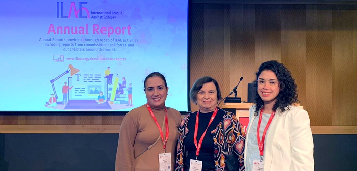
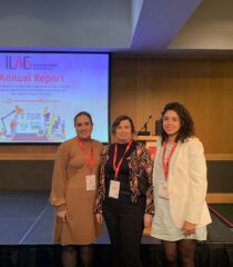
Os pesquisadores Fernando Cendes, Iscia Lopes-Cendes, Jaqueline Geraldis e Amanda Morato do Canto participaram do 35th International Epilepsy Congress (IEC), evento organizado pela International League Against Epilepsy (ILAE) e que este ano ocorreu entre os dias 02 e 06 de setembro na cidade de Dublin, na Irlanda. O evento contou com a participação de mais de 350 palestrantes de todo o mundo, distribuídos em mais de 120 sessões, abordando temáticas diversas sobre avanços científicos e médicos na epilepsia. Mais de 1300 trabalhos foram apresentados no total.

Fernando Cendes foi o speaker convidado para 02 sessões: uma palestra no sábado, dia 02, intitulada “Common epileptic pathologies: temporal epilepsy“, durante o curso “Neuroimaging in epilepsy – what the clinician should know“, e uma na quarta-feira, dia 06, com o tema “How to report and integrate neuroimaging findings of FCD“, durante o painel “A multi-layered and integrative classification scheme for FCD: the ILAE consensus classification update 2022“.

Iscia Lopes-Cendes apresentou o tema “Commonalities between multi-omics studies in basic and clinical epilepsy, do they translate?” durante o painel “Multi-omics in acquired epilepsies: beyond genes and transcripts“, que ocorreu no dia 05 de setembro.

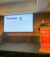
Amanda M. do Canto apresentou o trabalho “Proteomics analysis of synaptosomes of brain tissue from patients with pharmacoresistant mesial temporal lobe epilepsy” e Jaqueline Geraldis o estudo “Uncovering the multi-omic single-nuclei landscape of hippocampal sclerosis type I from patients with mesial temporal lobe epilepsy: clues into mechanism“. Veja detalhes sobre estes trabalhos ao final do post.

Confira a seguir fotos da participação dos pesquisadores do BRAINN no evento. Clique para abrir as imagens em alta resolução.


Detalhes sobre os trabalhos apresentados

Proteomics analysis of synaptosomes of brain tissue from patients with pharmacoresistant mesial temporal lobe epilepsy

A Morato do Canto1, J C Geraldis1, F Rogerio2, M Alvim3, C Yasuda3, E Ghizoni3, H Tedeschi3, M MacDonald4, F Cendes3, I Lopes-Cendes1

1University of Campinas, Department of Translational Medicine, Campinas, Brazil, 2University of Campinas, Department of Pathological Anatomy, Campinas, Brazil, 3University of Campinas, Department of Neurology, Campinas, Brazil, 4University of Pittsburgh, Department of Psychiatry, Pittsburgh, United States

Purpose: The study of synaptosomes can give clues about synaptic transmission and its abnormalities since they contain all the machinery involved in releasing, reuptake, and storing neurotransmitters. In addition, synaptic proteins may be therapeutic targets in many neurological diseases. Mesial temporal lobe epilepsy (MTLE) is the most common type of focal epilepsy in adults, with a high proportion of patients who do not respond to treatment with antiseizure medication. This study aims to characterize the proteome of synaptosomes isolated from the tissue of patients with pharmacoresistant MTLE.

Method: We analyzed the synaptosomes from brain tissue obtained from epilepsy surgery (hippocampus and anterior temporal lobe) of patients with pharmacoresistant MTLE (N=20 – 5 per group) and compared these with post-mortem control tissue (N=5). We classified the patients into four groups according to disease duration determined at the time of epilepsy surgery, 10, 20, 30, and 40 years. We isolated the synaptosomes, and proteomic data were acquired using an Orbitrap EclipseTM TribridTM (MacDonald Laboratory – University of Pittsburgh). We used the ProteomeDiscoverer and R software for bioinformatics analysis. We also performed a SynGO analysis to evaluate the identified biological classes of proteins.

Results: Overall, we identified 1,890 proteins and 7,521 peptides. We found differences in the protein content of the synaptosome of the four groups of patients. A preliminary analysis showed that most proteins identified in the synaptosome of patients belong to the presynaptic fraction, followed by the post-synaptic and the synaptic membrane.

Conclusion: Our study explores for the first time the protein content of the synaptosome in the brain tissue of patients with pharmacoresistant MTLE. As an initial finding, we identified differences in the proteomic content of these vesicles according to the duration of epilepsy. In addition, we expect to identify novel proteins that may play a role in disease mechanisms and pharmacoresistance.

Uncovering the multi-omic single-nuclei landscape of hippocampal sclerosis type I from patients with mesial temporal lobe epilepsy: clues into mechanism

J Geraldis1,2, D Veiga1,2, D C.F. Bruno1,2, F Rogerio3, A Gaigneaux4, K Grzyb5, M K.M. Alvim1,6, C L. Yasuda1,6, F Cendes1,6, T Sauter4, L Sinkkonen4, I Lopes-Cendes1,2

1Brazilian Institute of Neuroscience and Neurotechnology (BRAINN), Campinas, Brazil, 2Department of Translational Medicine; School of Medical Sciences, University of Campinas, Campinas, Brazil, 3Department of Pathology; School of Medical Sciences, University of Campinas, Campinas, Brazil, 4Department of Life Sciences and Medicine; Faculty of Science, Technology and Medicine, University of Luxembourg, Luxembourg, Luxembourg, 5University of Luxembourg / Luxembourg Centre for Systems Biomedicine, Luxembourg, Luxembourg, 6Department of Neurology; School of Medical Sciences, University of Campinas, Campinas, Brazil


Purpose: We aim to construct a compressive map of molecular interactions occurring in tissue obtained by epilepsy surgery of patients with pharmacoresistant mesial temporal lobe epilepsy with hippocampal sclerosis (MTLE+HS) using single-cell/nuclei omics techniques. Furthermore, with this approach, we can identify the molecular pathways activated in different cell populations and understand better how the cells’ epigenome landscape may change over time.

Method: We selected 14 samples from patients with MTLE+HS and classified them as type I hippocampal sclerosis by post-surgical pathological examination. Samples were divided into three groups according to disease duration (DS): Group 1: DS≤25 years (n=3); Group 2: 26 years≤DS≤39 years (n=5); Group 3: DS≥40 years (n=6). In addition, neuronal cell nuclei were isolated, resulting in 28 multi-omic libraries (14 Single Nuclei ATAC + 14 Single Nuclei Gene Expression; 10X Genomics Multiome Protocol).

Results: To date, our results identified 16 clusters of different cell populations, with a median of 1.038 GEX/cell and 96.090 peaks corresponding to genomic regulatory regions of open chromatin. Furthermore, we obtained a total of 75,445 cells aggregating all samples. The identification of different cell types, expression gene patterns, and regulatory elements is underway.

Conclusion: To our knowledge, this is the first study applying a single-nuclei multi-omic approach focusing on studying molecular changes occurring over time in tissue from patients with MTLE+HS. We obtained high-quality sequencing data from more than 70.000 single cells and identified 16 different cell sub-populations from the sclerotic hippocampi of patients. Our work may contribute to a better understanding of disease changes over time, especially in identifying key activated molecular mechanisms, changes in gene expression regulation, and the overall epigenome landscape at different disease time points in patients with MTLE-HS.


Website oficial do evento


Compartilhe! / Share this!